Your cart is currently empty!

In recent years, antibody-oligonucleotide conjugates have emerged as indispensable tools in scientific research, enabling unparalleled capabilities in multiplexing, detection, and data integration. From single-cell multi-omics to high-throughput imaging and advanced diagnostics, antibody-oligo conjugates are reshaping the way researchers explore complex biological systems.
Here we highlight some recent work from our customers showcasing novel applications and research methodologies that leverage oYo-Link® antibody-oligo conjugates.
1. Cassini: A Streamlined and Scalable Method for In Situ Profiling of RNA and Protein
At the end of 2024 we saw the authors of this research present Cassini, a cost-effective method for multiplexed mRNA and protein analysis in intact tissue.
Utilizing rolling circle amplification (RCA), Cassini achieves robust signal amplification, enabling the detection of multiple targets within 20 minutes. This technique overcomes limitations of existing methods like single-molecule FISH by providing higher multiplexing capabilities while remaining accessible and scalable. Additionally, Cassini supports simultaneous immunostaining with fluorophore-conjugated or DNA-barcoded antibodies, making it a versatile tool for spatial genomics applications.
The researchers used oYo-Link® Oligo to link RNA-targeting probes with DNA-barcoded antibodies – anti-GFAP and anti-Neun antibodies – enabling simultaneous immunostaining and serial feature detection in under 20 minutes.
Read more about this research:
Cassini: Streamlined and Scalable Method for in situ profiling of RNA and Protein
2. BDEVS: Ultra-Sensitive Extracellular Vesicle Profiling
In November 2024 we saw a study presenting the BDEVS platform (Agarose Bead-based Digital Single Molecule-Single EV Sorting) for the high-throughput, multiplexed, and ultra-sensitive profiling of individual extracellular vesicles (EVs) directly in plasma.
Unlike traditional methods, BDEVS delivers single-molecule sensitivity and moderate multiplexing (demonstrated with 3-plex) while maintaining the high throughput required to analyze extracellular vesicles (EVs) directly in human plasma, processing tens of thousands of EVs per minute.
The platform integrates rolling circle amplification (RCA) of EV surface proteins, which are cleaved from single EVs and amplified within agarose droplets. This approach overcomes challenges such as steric hindrance and non-specific binding that have affected earlier methods. The amplified signals are then analyzed and sorted using flow cytometry, enabling precise quantification of multiple proteins on individual EVs. BDEVS has demonstrated high-throughput profiling of key immune markers like PD-L1, CD155, and the melanoma tumor marker TYRP-1, offering unprecedented resolution for analyzing tumor-immune interactions and detecting rare EV subpopulations in complex clinical specimens.
The researchers used oYo-Link® Oligo to conjugate DNA oligomers to the Fc region of antibodies, ensuring consistent primer attachment and enabling precise quantification of biomarkers on individual EVs.
Read more about BDEVS here:
High-throughput, multiplexed quantification, and sorting of single EVs at single-molecule level
3. DNA-based AND logic gate as a molecular precision tool
In a working paper in ChemRxiv, the authors introduce a DNA-based molecular logic AND gate, designed to recognize the simultaneous presence of two specific biomolecules—such as membrane proteins PD-L1 and CD3—by utilizing corresponding antibody-oligonucleotide conjugates. This dual-recognition mechanism ensures that the gate activates only when both target proteins are present, enhancing specificity in applications like diagnostics and targeted therapy.
In this system, each antibody is linked to a DNA strand that serves as an input signal. When both antibodies bind to their respective target proteins embedded in lipid nanodiscs (modeling cell membranes), the proximity of the DNA strands facilitates a hybridization event, effectively “closing” the logic gate. This activation can trigger subsequent actions, such as the binding of DNA-tagged gold nanorods, which could be employed for photothermal therapy. This approach offers a molecular precision tool capable of processing multiple input signals to produce a specific output, paving the way for high-specificity diagnostics and therapeutic interventions.
The researchers used oYo-Link® Oligo to link input DNA strands to monoclonal antibodies, enabling targeted and specific interaction with the desired antigens, integrating seamlessly into the logic gate construct.
Read more about DNA-based AND logic gate here:
4. cycleHCR: Expanding Spatial Omics Capabilities
Spatial biology takes another leap forward with cycleHCR, a technique leveraging multicycle DNA barcoding and hybridization chain reaction (HCR) to overcome the limitations of traditional fluorescence microscopy, that constrain comprehensive spatial analysis in biological specimens.
cycleHCR facilitates high-specificity, single-shot imaging per target for RNA and protein species within thick specimens, mitigating the molecular crowding issues encountered with other imaging-based spatial omics techniques.
To harness the high specificity of cycleHCR for multiplex protein imaging, the researchers used oYo-Link® Oligo to form an antibody complex that simultaneously recognizes the target and anchors a cycleHCR barcode within a polyacrylamide gel for imaging readout.
Read more about cycleHCR here:
5. FLASH-PAINT: Super-Resolution Imaging
Research published in Cell in March 2024 introduces FLASH-PAINT, a method for rapid, multiplexed super-resolution microscopy capable of imaging a nearly unlimited number of molecular targets. Traditional super-resolution fluorescence microscopy is limited to detecting a few targets at a time, while methods that rely on sequential imaging rounds struggle with low throughput. FLASH-PAINT addresses these limitations by combining fluorogenic labeling with transient adapter-mediated switching, enabling high-throughput imaging with exceptional resolution.
The versatility of FLASH-PAINT is demonstrated through several applications: mapping nine proteins in a single mammalian cell, studying the functional organization of primary cilia, analyzing changes in the proximity of thirteen molecular targets in Golgi stacks, and investigating inter-organelle contacts in 3D super-resolution. This technique significantly enhances the ability to map the spatial relationships of diverse molecules within cells, offering powerful insights into cellular function and organization.
oYo-Link Oligo was used to label primary antibodies for FLASH-PAINT imaging of normal and bulbous-tip cilia in RPE-pHSmo cells.
Read more about FLASH-PAINT here:
Why our customers choose oYo-Link® for their oligo conjugations
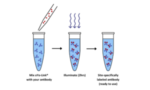
AlphaThera’s oYo-Link® Oligo technology enables fast, simple and site specific antibody labeling with custom oligos, using only the amount of antibody you need for your experiment, and without a large excess of oligo.
Unlike other oligonucleotide conjugation products available, oYo-Link allows site specific, covalent labeling of antibodies in the Fc region. This guarantees there will be no interference with antibody binding sites, as well as a homogenous mixture of antibody-oligo conjugates with 1 to 2 oligonucleotides attached per antibody. Therefore, oYo-Link Oligo gives scientists greater control and precision in their experiments, removing any impact of decreased assay performance associated with traditional heterogeneous labeling products.
Advantages of oYo-Link® for antibody-oligonucleotide conjugation:
- Label as little as 1ug of antibody at a time – reduce overall cost and minimize wasted materials
- Custom oligos supported, up to 80bp or longer, for ssDNA or dsDNA.
- Easy and rapid oligo-antibody labeling with less than 30 seconds hands-on time.
- Site specific labeling in the Fc region, so no loss in antibody functionality or batch-to-batch variability.
- Oligos are all HPLC or PAGE grade ensuring confidence in downstream assays.
- Label diluted antibody – as low as 50 μg/mL – saving time as antibodies do not need to be concentrated prior to labeling.
- No need for purification prior to conjugation – compatible with nearly all buffers, even those containing Tris and albumin.
Download our brochure to find out more about oYo-Link® Oligo, or don’t hesitate to get in touch with any technical questions.